


Usefulness of MRI for the differential diagnosis of suture recurrence and suture granuloma after pulmonary resection for lung cancer
MRI (DWI) can assess new suture lesions after pulmonary resection of lung cancer (Fig.26, 27).
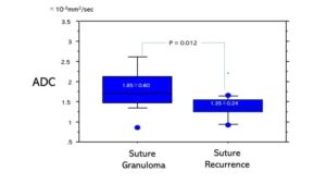 Fig. 26 Usefullness of MRI (DWI) for suture lesions after lung cancer resection Difference of ADC value between suture granuloma and suture recurrence of lung cancer. ADC value (1.35±0.24 x10⁻³ mm2/sec) of suture recurrence was significantly lower than that (1.85±0.60 x10⁻³ mm2/sec) of suture granuloma (P=0.012). Usuda K, et al. Transl Oncol. 2021;14(2): 100992
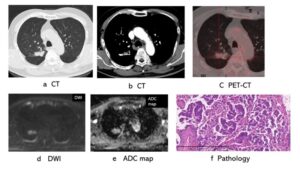 Fig. 27 A 67-year-old male underwent partial resection of right upper lobe for adenocarcinoma. One year and 10 months after the resection, a new shadow at the suture developed and a right upper lobectomy was performed and it revealed a suture recurrence of the adenocarcinoma. a, b: CT showed a new lesion at the suture. c: PET-CT showed a slight accumulation (SUVmax 4.1) of FDG. d: Diffusion detectability score was ”moderately detectable.” e: ADC was 1.464 x 10⁻³mm2/sec on ADC map. f. HE stain of the suture recurrence (adenocarcinoma)
Usuda K, et al. Differentiation between suture recurrence and suture granuloma after pulmonary resection for lung cancer by diffusion-weighted magnetic resonance imaging or FDG-PET /CT. Transl Oncol. 2021;14(2): 100992.doi: 10.1016/j.tranon.2020.100992.