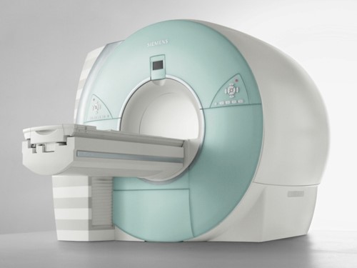


17. DWIBS ( Diffusion-weighted whole-body imaging with background suppression: Whole-body DWI)
On Feb 26, 2022 Nippon Television program (The most desirable lecture in the world !! Japanese great medical care !!) reported that DWIBS is the ultimate examination which can detect every type of cancer in the whole body by only one DWIBS scan. Surprisingly, a patient is able to eat before a DWIBS scan, unlike other diagnostic methods available.
The research group of Dr. Taro Takahara and Professor Dr. Yutaka Imai of Tokai University have developed an epoch-making method, diffusion-weighted whole-body imaging with background suppression (DWIBS) which searched a whole body with one MRI (DWI) exploration in 2004. The diagnostic ability of PET-CT and that of DWIBS are similar. Some diagnostic abilities are better in DWIBS than in PET-CT, others are better in PET-CT than DWIBS. However, patients cannot receive a PET-CT more than once a year on health insurance because it is expensive, approximately 30,000 yen with a 30% deductible. On the other hand, DWIBS is an MR imaging so we do not need a contrast medium. Therefore, a patient can get an MRI once every one to three months on health insurance.
We presented the good points of DWIBS compared to PET-CT (Figure 16). Also, we showed a comparison between DWIBS and PET-CT (Figure 17).

 One of the most famous journals in American radiology is “Radiology”. Mark L. Schiebler,M.D. as EDITORIAL of Radiology in 2016 cited our paper of whole-body DW MRI (DWIBS) for lung cancer. He mentioned that if the diagnostic ability of whole-body DW MRI (DWIBS) is proved to be equivalent to PET-CT for clinical staging of lung cancer while also reducing medical cots it will ultimately replace PET-CT in the future. Furthermore he predicted that only whole-body DW MRI (DWIBS) will be utilized for the diagnosis and staging of lung cancer.
Mark L. Schiebler,M.D. Can solitary pulmonary nodules be accurately characterized with diffusion-weighted MRI? Radiology 2019; 290:535–536 https://doi.org/10.1148/radiol.2018182442
(Quoted sentence) There is a single report by Usuda et al. that shows that DW MRI can be used to adequately stage NSCLC. In their study of 67 patients with NSCLC, PET/CT plus brain MRI showed a pathologic staging accuracy of 0.69, while in the same group, whole-body DW MRI had a pathologic staging accuracy of 0 .75. This data (8) clearly points to a need for an adequately powered prospective randomized trial to help definitively answer this question. Specifically, if whole-body DW MRI can be shown to have equipoise with 18F-FDG PET for the clinical staging of NSCLC, this would reduce the costs of patient work-up because 18F-FDG PET would no longer be needed. Perhaps in the near future, only whole-body DW MRI will be needed for clinical staging in patients with a new diagnosis of NSCLC.
(Quoted paper) Usuda K, et al. Diagnostic Performance of Whole-Body Diffusion-Weighted Imaging Compared to PET-CT Plus Brain MRI in Staging Clinically Resectable Lung Cancer. Asian Pac J Cancer Prev. 2016; 17: 2775-2780.
18. DWIBS for detection of recurrence and metastasis after surgery of lung cancer (Figure 58-62)
We reported that DWIBS could detect recurrence and metastasis with a better accuracy after surgery of lung cancer.
Usuda K, Iwai S, Yamagata A, Iijima1 Y, Motono N, Matoba M, Doai M, Yamada S, Ueda Y, Hirata K, Uramoto H. Diffusion‐weighted whole‐body imaging with background suppression (DWIBS) is effective and economical for detection of metastasis or recurrence of lung cancer. Thoracic cancer 2021:12 (5):676 – 684. doi:10.1111/1759-7714.13820
Background: Diffusion-weighted whole-body imaging with background suppression (DWIBS) is used for the diagnosis and staging of cancers. The medical cost of an MR examination including DWIBS is $123, which is 80% less expensive than the cost ($798) of F18-fluorodeoxyglucose positron emission tomography/computed tomography (FDG-PET/CT) examination. Methods: This study examined the efficacy of DWIBS for relapses after lung cancer resection. A total of 55 patients who had pulmonary resection of lung cancer, postoperative computed tomography (CT) every six months, and DWIBS and FDG-PET/CT (every year) were enrolled in this study. If a metastatic lesion was detected on CT scan, DWIBS and FDG-PET/CT were also used. Results: A total of 55 patients who underwent pulmonary resections for lung cancer, and had CT, DWIBS and FDG-PET/CT examination during follow-up after pulmonary resection were enrolled in this study. Lung cancer in 32 patients relapsed. Postoperative radiographic examinations revealed pulmonary metastases in 17 patients, bone metastases in seven, liver metastases in five, lymph node metastases in five, pleural metastases in four, metastases to the chest wall in two, brain metastases in two, adrenal gland metastasis in one, and renal metastasis in one. The mean apparent diffusion coefficient (ADC) value of the relapse was 0.9 to 1.70 × 10⁻³ mm2/s. The accuracy 0.98 (54/55) of DWIBS for detecting multiple metastatic lesions was likely to be higher than 0.94 (52/55) of CT or 0.94 (52/55) of FDG-PET/CT, but there were no significant differences. Conclusions: DWIBS can detect multiple metastatic lesions throughout the entire body and differentiate malignancy from benignity in only one examination. DWIBS has benefits of diagnostic accuracy and is less expensive in medical costs for the detection of a relapse. DWIBS could potentially replace FDG-PET/CT after lung cancer resection.