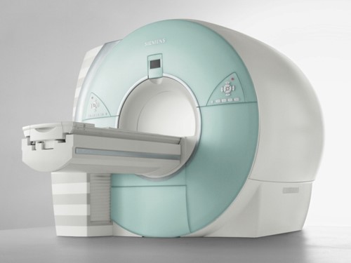
Characteristics of basic findings of MRI
Various images taken with an MRI have different characteristic and represents different information. It is valuable to know the characteristics of each type of MRI scan to better understand the MRI images of lung cancer and other malignant tumors.
MRI-DWI imagining has become an essential tool in diagnosing early stage cerebral infarction.
It is also interesting that diffusion weighted imaging has become valuable in the diagnosis of many malignant tumors.
Characteristics of MRI are as follows:
- No radiation exposure
- Detailed images of bones, soft tissue, ligaments, organs and tumors.
- A variety of cross sections can be obtained and the qualitative diagnosis of lesions can be detected.
- The angiograms can be obtained without using a contrast medium.
Characteristics based on the sequence of the MRI are as follows:
- T1 WI (weighted imaging):Water, humoral components, and cysts look black. Fat and contrast mediums look white. In T1 WI, anatomical structures are easy to recognize.
- T2 WI (weighted imaging):Not only the adipose tissue but also the humoral components and cysts look white. Tumors look moderately white. Bleeding looks black. A lesion in the acute phase is easy to recognize with T2 WI.
- Diffusion-Weighted Imaging (DWI): DWI is used to detect the movement of water molecules. Due to this ability to accurately measure the movement of water molecules in the body DWI is very effective for diagnosing the acute phase of cerebral infarctions.
- DWI creates images based on the diffusion motion of water molecules. Lesions such as tumors, infarctions, and inflammation have less diffusion activity in their DWI images.
- Originally, DWI was mostly used for diagnosing cerebral infarctions, but recently DWI is being used to scan the whole body to detect and diagnose malignant tumors. MRI can detect smaller tumors because of its higher spatial resolution. Due to DWI’s ability to detect decreased diffusion it can detect cancers in the early stages.
- DWI can detect smaller tumors such as liver, kidney, and bladder cancers that were difficult to detect in other modalities of examinations.
- One drawback of DWI is that DWI shows decreased diffusion (higher signals) not only for malignant tumors, but also for normal tissues such as spleen, ovary, lymph nodes, inflammation, abscesses and benign tumors. It is necessary to pay attention and be mindful of false positive results.
- MR angiography:MRI angiography without a contrast medium can show only blood vessels, and it is easy to detect obstruction and neovascularity of blood vessels.
Figure 63. Changes over time of the findings of the MRI sequences in cerebral infarction


Interpretation of the MRI image
- A brain lesion with a high DWI signal and ADC map with a low signal is highly suspected to be a cerebral infarction
- A brain lesion with a high DWI signal and ADC map with a high or normal signal is not suspected to be a cerebral infarction.
- Even a high DWI signal may not be a cerebral infarction.
- A T2 shine through is where pathologic abnormalities cause a long T2 decay time in some benign tissue. An old cerebral infarction sometimes shows a high DWI signal which was not caused by restricted diffusion but by a higher T2 WI signal.
- We have to refer to the ADC map to discriminate whether it is a cerebral infarction or not.
Points of the interpretation
- If the ADC map presents a low signal then there is the possibility of a cerebral infarction.
- The problem with DWI is the differentiation between a malignant tumor and an abscess. Even, in that case, we can confirm the diagnosis from the signal pattern in reference to the ADC map and T2 WI.
With MRI, the combinations of several image sequences can make qualitative diagnoses of cerebral infarctions and malignant tumors.


